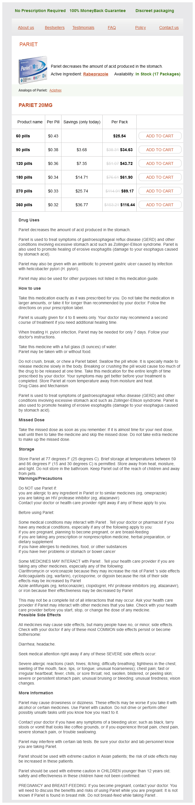
Pariet dosages: 20 mg
Pariet packs: 60 pills, 90 pills, 120 pills, 180 pills, 270 pills, 360 pills

Discount pariet 20 mg free shipping
These include: � Spleen can sometimes decrease in dimension and launch as a lot as one hundred ml of blood into the reservoir of circulation. Conduits the systemic veins carry blood from the tissues to the proper atrium, and the pulmonary veins gather blood from the lungs and return it to the left atrium. After blood loss from an exterior or internal damage, reflex enhance in sympathetic discharge produces contraction of the graceful muscles within the walls of the veins. As a result of venular contraction, there occurs a lower of their capability leading to increased venous return to coronary heart. In this fashion, veins assist to preserve cardiac output by sustaining normal venous return despite blood loss. Venous pressure and flow the venous system is a low-resistance, low-pressure and highly distensible a half of the vascular system. At normal pressures, the veins are approximately 20 instances extra compliant than the arteries. Central venous pressure Central venous pressure refers to the stress in the best atrium as a outcome of all of the systemic veins open into the best atrium. The proper atrial stress can lower to as low as -3 to -5 mmHg when the guts (right atrium) is pumping with vigour or when venous return is significantly depressed. Therefore, massive veins do offer considerable resistance to blood move and thus stress in the peripheral veins is often higher (4�7 mmHg) than that of the best atrial strain. This produces a strain gradient which propels the venous blood towards the center (venous return). Gravitational hydrostatic stress happens within the vascular system due to the weight of the blood within the vessels. Peripheral venous stress, like arterial stress, is affected by this gravitational hydrostatic stress. Therefore, if the venous valves are incompetent, as in varicose veins, it ends in venous pooling, i. Therefore, neck veins completely collapse due to atmospheric strain on the skin of neck, and pressure contained in the vein nearly remains zero. Because of this cause, if a dural sinus is opened throughout a neurosurgical procedure with the patient seated, air is sucked into the sinus, leading to air embolism. Like arterioles, myogenic tone of the veins induced by sympathetic constrictor nerves helps to regulate the capability of vascular system. For instance, sudden change of posture from lying down to standing position results in peripheral pooling of the blood in veins of legs and ft as a result of effect of gravity. Therefore, venous return to coronary heart decreases, systemic blood pressure falls and may trigger dizziness. However, normally, the compensatory mechanism operated by way of baroreceptor reflex prevents any fall in blood stress (see page 335). Increase in venous resistance due to compression of peripheral veins inside or without causes an increase in peripheral venous stress. Peripheral venous stress, to an excellent extent, depends upon the central venous strain. Common causes related to rise in proper atrial strain are: � Increased blood quantity (massive blood transfusion, � Heart failure and � Arteriolar dilation decreases peripheral resistance inflicting fast flow of blood from arterioles thus increasing the central venous pressure. The venous strain in ft is roughly +90 mmHg in a standing position due to hydrostatic stress effect. However, actions of legs and muscle contractions (muscle pump or venous pump) squeeze the blood out of veins; the valves within the veins are arranged in such a way that the course of blood can solely be towards the heart. Measurement of peripheral and central venous stress � Clinical evaluation of venous stress is made by observing the diploma of distension of neck veins. When right atrial pressure is increased up to 10 mmHg, the lower neck veins start to protrude in sitting place (in normal person, in this position, the neck veins are by no means distended). Venous flow and venous return As we all know, the blood flows within the veins in direction of the heart due to a stress gradient which exists between the best atrial strain (0 mmHg) and the peripheral veins (6�7 mmHg). The velocity of blood move within the veins will increase with enhance within the size of vein. Blood stress Definitions (terminology) Blood pressure Blood pressure is the lateral pressure exerted by the flowing blood on the walls of the vessels. Without any additional qualification, the time period blood pressure denotes the arterial pressure.
Syndromes
- Lack of alertness (stupor)
- Localized swelling
- Sadness
- The surgeon uses the tools to remove the urachal tube and close off the bladder and area where the tube connects to the umbilicus.
- Migraine - resources
- Diabetes
- Heart failure
- Dehydration
- Circular marks around the wrists or ankles (signs of twisting or tying up)
- The ovaries stop making the hormones estrogen and progesterone.
Buy pariet cheap online
For the aim of representation, the cerebellar cortex has been divided into three zones: � Vermal zone, i. There is a double representation on the superior surface (anterior area) and on the inferior surface (posterior area). Axial parts of the body lie within the vermis, whereas limbs and facial region lie in the intermediate zone of cerebellar cortex. In this area, the physique representation is a bilateral projection, less outlined and is erect. Stimulation of those areas produces movements in elements of the physique that correspond roughly to these from which sensory impulses are received. These areas receive signals entirely and completely from the cerebral cortex, and premotor areas of frontal cortex, somatosensory and sensory affiliation areas of parietal cortex. These connections play an necessary position in planning and co-ordinating fast sequential muscular activities of the physique. The primary afferents that converge to kind mossy fibres and climbing fibres to the cerebellum are summarized in Table 10. The fibres originate from dentate nucleus, which is recent in origin and most well developed in man and so the red nucleus, thalamus and cerebral cortex of opposite side. Cerebellovestibular fibres cross via the inferior cerebellar peduncle to vestibular nuclei. These efferents control anterior horn cells of the spinal wire via vestibulospinal tract. Some fibres from the cerebellum additionally attain the nucleus of oculomotor nerve and the tectum. Functions of cerebellum Functionally, cerebellum has been divided into three divisions: � Vestibulocerebellum, � Spinocerebellum and � Corticocerebellum. These three functional divisions play necessary role in following completely different capabilities: A. Control of physique posture and equilibrium Vestibulocerebellum, which includes flocculonodular lobe as its principal part and nucleus fastigii (as its effector nucleus) and vermal region of the cerebellum, are concerned with management of physique posture and equilibrium. Afferents to cerebellum concerned with the control of physique posture and equilibrium include: � Vestibulocerebellar tracts which carry enter from vestibular nuclei, which convey afferents from the macula of saccule and utricle for static equilibrium and from the ampullary crests of semicircular ducts for kinetic equilibrium. The flocculonodular lobe and fastigial nuclei project output fibres through inferior peduncle to vestibular and reticular nuclei of brainstem. The vermal cerebellum sends again the information to spinal wire indirectly via fastigial nuclei. The efferents from the cerebellum affect the spinal motor neurons to keep the body posture upright by way of the vestibulospinal and reticulospinal tracts, and regulate the position of eyes in relation to movements of the top by connecting motor nuclei of extraocular muscular tissues (3rd, 4th and sixth cranial nerves) by way of medial longitudinal fasciculus. It is essential to notice that the cerebellum does needed corrections for sustaining posture and equilibrium, without participation of conscious will and that the corrections made are highly easy and precise. Control of muscle tone and stretch reflexes Spinocerebellum, which incorporates entire anterior lobe besides lingula and a few components of posterior lobe (pyramis, uvula and paraflocculus) as its principal elements and nucleus globossus and nucleus emboliformis as its effector nuclei, is especially concerned with management of muscle tone and anticipatory adjustment of muscle contraction during motion. Afferents spinocerebellar, cuneocerebellar and olivocerebellar tracts carry proprioceptive and tactile inputs from the limbs, trunk, neck and other elements of the physique. Spinocerebellum additionally receives auditory and visual impulses through tectocerebellar tract. The spinocerebellum is projected into the cerebellar nuclei-fastigii, emboliformis and globossus, Fibres from these nuclei pass via fastigiobulbar, cerebelloreticular and cerebello-olivary tracts and finally, to relay to the and motor neurons via the reticulospinal and olivospinal tracts. The motor neurons reflexly modify the activity of motor neurons and thus regulate the muscle tone. Thus, cerebellum types an important site of linkage of � techniques responsible for muscle tone. Temporary suppression of anterior lobe exercise by surface cooling abolishes discharge from the motor neurons, leading to hypotonia and disturbance in posture. Therefore, lesions of cerebellum are associated not with paralysis however with disturbances within the smoothness of movements. Control of actions by cerebellum consists of regulation of time, rate, range (extent), pressure and path of muscular exercise. Control of voluntary movements includes co-ordination of activity of the muscle tissue involved in voluntary movements which could be categorised as: � Agonists or prime movers, whose contraction is basically answerable for the movement of the part, � Antagonists are those which oppose the prime movers, � Synergists are those which assist the prime movers and reduce pointless involvements to a minimal and � Fixation muscular tissues, whose contraction causes the fixation of neighbouring joints and maintain the limbs or physique ready applicable for finishing up the actual movement. Pathway of management of voluntary movements Corticocerebellum also known as cerebrocerebellum is mainly concerned with integration and regulation of well-co-ordinated muscular exercise (though different components of cerebellum also work in close co-operation on this task).
Pariet 20 mg order without a prescription
Lymphatic circulation which is disposed in parallel to the circulation of blood could be considered a third type of circulation. Some tissue fluid enters the lymphatic channels as lymph, which is finally drained into the venous system by way of the thoracic lymphatic duct and the best lymphatic duct. Systemic vascular tree the arrangement of systemic circulation permits extensive variations in regional distribution of blood. For descriptive purposes and from a functional viewpoint, the systemic vascular tree could be divided into the following types of blood vessels: � Large elastic arteries (Windkessel vessels) embody aorta and its primary branches corresponding to carotid, iliac and axillary arteries, � Large muscular arteries (distribution vessels) which embody a lot of the arteries of the body. Structure of blood vessels Structural traits General structural characteristics Histologically, walls of most of the blood vessels except the capillaries encompass three coats. In giant arteries, from inside-out, it consists of: � Endothelial lining, which could be very easy and silky, and consists of single layer of cells. On the surface, tunica media is restricted by a membrane shaped by elastic fibres referred to as the external elastic lamina. Important points to be noted are: � Fibrous parts within the intima and adventitia (mainly collagen) run longitudinally. Specific structural characteristics � Large (elastic) arteries, in their tunica media, have dominant elastic tissue which provides them property of distensibility and elastic recoil. In these arteries, the elastic tissue, both in intima and media, is far less and thus the proportion of smooth muscles increases. Their media consists of a thick layer of clean muscle tissue and so they have a relatively slim lumen. A cuff of smooth muscle cells known as the precapillary sphincter surrounds the origin of capillaries in some area. It is fashioned essentially by endothelial cells that are lined on the outside by a basal lamina (glycoproteins), branching perivascular cells referred to as pericytes. Essential characteristics Essential characteristics of blood vessels like lumen diameter, wall thickness, approximate whole cross-sectional area and percentage of blood quantity contained are shown in Table four. Arrangement in tunica media of the vessels Vascular clean muscle is present in all segments of vascular tree, except the capillaries. Innervational characteristics � Smooth muscular tissues of the blood vessels are innervated by sympathetic fibres. Therefore, noradrenaline causes contraction of muscle fibres leading to vasoconstriction. Thus, sympathetic stimulation elevates blood strain which tends to stay for a while. Haemodynamics Haemodynamics, which refers to research of blood circulate in various segments of the vascular system, could be mentioned under the next headings: � General ideas governing (factors affecting) blood flow, � Circulation time, � Types of blood flow, � Measurement of blood move, � Distribution of blood circulate to various areas of the body and � Regulation of blood move in several conditions. General rules governing (factors affecting) blood move Flow- pressure-�resistance relationship Relationship between circulate of a fluid with the pressure and resistance supplied to it through a inflexible tube was studied by a French physiologist Poiseuille in 1842. Hagen, a recent of Poiseuille, labored additional with the issue and discovered the mathematical expression. In different phrases, fluid at all times flows from an area of excessive strain (P1) to considered one of decrease stress (P2), and price of flow (Q) is determined by the strain gradient (P1 - P2), i. This is well comprehensible, as every phase of the tube is providing resistance to the flow; therefore, longer the size, larger will be the complete resistance offered. According to mathematical calculation in principles of physics, resistance (R) is represented by eight L/r4. Note the progressive lower in strain from the left ventricle through the systemic circulation till blood enters the right ventricle. It is essential to notice that the best pressure drop happens in the arterioles, which characterize the best resistance section of the systemic circulation. The relationship between move of blood and pressure in the blood vessels is nevertheless not linear: � In distensible vessels. This myogenic contractile response to stretch offsets the elastic impact and so the flow in these vessels turns into lower than the inflexible tubes, i. So, a certain quantity of intramural stress is a should to counteract the tissue strain and thus to hold the vessels patent and to preserve the blood flow. Equilibrium of things at crucial closing strain � In general, vasomotor tone of the blood vessels tries to constrict the vessels. This tone is elevated by sympathetic stimulation and decreased by sympathetic inhibition.
Discount pariet 20 mg online
The ossification is carried out by osteoblasts which enter the central part of the cartilaginous model. At in regards to the time of start, developing bone consists of the bony diaphysis shaped by extension of primary centre for ossification and cartilaginous ends. At varying instances after birth, secondary centres of endochondral ossification seem in the cartilages forming the ends of bones. The bone increases in length as this plate lays down new bone on the tip of shaft. The bone increases in size as long as the epiphyseal plates remain separated from diaphysis (shaft). The development of the bone stops when the epiphysis fuses with the diaphysis (epiphyseal closure). The epiphyseal closure occurs in an orderly temporal sequence, the final epiphysis closing after the puberty. The normal age at which the epiphysis closes in different bones of the body is well known, and the age of a younger particular person may be determined by looking at the open and closed epiphysis in radiograph of the skeleton. Even after bone development has ceased, the calcium turnover operate of bone is most active in the metaphysis which acts as a storehouse of calcium. Bone formation Bone formation is carried out by active osteoblasts, which is why these are additionally called bone-forming cells. The process of bone formation may be thought of in following steps: � Formation of bone lamellae, � Formation of trabecular bone and � Formation of compact bone. It consists of two primary processes: osteoid formation and mineralization of bone matrix. The collagen polymerizes to type collagen fibres which then swell up and can now not be seen distinctly. Bone matrix mineralization Soon after formation of osteoid, the method of bone matrix mineralization begins. It happens in two phases: an initial gradual strategy of initiation of mineralization followed by rapid mineralization process. The bone matrix is surrounded by a metastatic solution of calcium and phosphate ions (solution by which focus of Ca2+ and Po43- exceeds the solubility product of the salt but precipitation is inhibited by certain inhibitors like pyrophosphate). For enucleation to happen, pyrophosphate has to be cleaved into inorganic phosphate by alkaline phosphatase which additionally has activity of pyrophosphatase. The strategy of mineralization greatly relies upon upon the calcium � phosphate ion product in extracellular fluid. Thereafter, hydroxide and bicarbonate ions are progressively added to the mineral combination, and mature hydroxyapatite crystals are slowly shaped. In this fashion, numerous lamellae are laid down one over one other and these lamellae together type a trabecula of bone, but many such trabeculae constitute the trabecular or cancellous bone. Within every lamella, mineral fluid containing channels, called canaliculi, traverse the mineralized bone. Through these channels, the interior osteocytes stay connected with floor lining cells and with other osteocytes through syncytial cell processes. This system of interconnected cells fashioned by osteocytes and osteoblasts spreads over all the bone surfaces besides small floor area adjacent to osteoclasts. A small quantity of fluid known as the bone fluid is current between the bone and osteocytic membrane. This arrangement permits transfer of calcium from the enormous floor space of the inside to the outside of the bone models, and then into the extracellular fluid. This switch course of, which is carried out by the osteocytes, is called osteocytic osteolysis. Conversion of trabecular bone to compact bone All newly fashioned bone is cancellous. In bone resorption, destruction of entire matrix of bone occurs leading to diminished bone mass.
Pariet 20 mg with mastercard
On reaching the appropriate degree of the spinal cord, the fibres of this tract cross the midline (through the anterior white commissure) to attain gray matter on the opposite facet of the twine and terminate in a way similar to that of the fibres of lateral corticospinal tract. Thus, the corticospinal fibres of each the lateral as nicely as anterior tracts ultimately join the cerebral cortex of one facet with ventral horn cells in opposite half of spinal wire. Salient features of nerve fibres of corticospinal tracts � Fibres of the corticospinal tract are unmyelinated at delivery. The cerebral cortex controls voluntary fine-skilled actions of the physique via the corticospinal tracts. Interruption of the tract anyplace in its course results in paralysis of the muscles involved. As the fibres are intently packed in their course via the inner capsule and brainstem, small lesions right here could cause widespread paralysis. Extrapyramidal tracts the descending tracts of spinal cord other than the pyramidal tracts are collectively known as extrapyramidal tracts. These embrace: � Rubrospinal tract, � Vestibulospinal tract, � Reticulospinal tract, � Tectospinal tract, � Olivospinal tract and � Medial longitudinal fasciculus. This tract arises from the large cells (nucleus magnocellularis) or pink nucleus in midbrain. After arising from the purple nucleus, the fibres of this tract cross to reverse side in the lower a half of segmental of midbrain (ventral segmental decussation). This tract reveals facilitatory influence on the flexor muscular tissues and inhibitory influence on the extensor muscle tissue of the body. The corticorubro-spinal tract thus formed could act as an alternate route of pyramidal system to exert affect on lower motor neurons. In humans, the red nucleus is comparatively small and the rubrospinal tract reaches only the higher three cervical segments of the spinal wire. Fibres to cervical segments arise from the cranioventral half, those to thoracic segments from the central part and those to lumbosacral segments from the dorsocaudal a half of lateral vestibular nucleus. Vestibular nucleus receives afferents from vestibular apparatus primarily from utricles. This pathway is principally concerned with adjustment of postural muscle tissue to linear acceleratory displacements of the body. Lateral vestibulospinal tract mainly facilitates exercise of extensor muscle tissue and inhibits the activity of flexor muscular tissues in association with the upkeep of stability. This tract descends through the anterior funiculus (within the sulcomarginal fasciculus). This a half of the vestibular nucleus receives indicators from the vestibular apparatus mainly from the semicircular canals. Functionally, medial vestibulospinal tract is the donor connection of medial longitudinal fasciculus. This tract supplies a reflex pathway for movements of head, neck and eyes in response to visible and auditory stimuli. Reticulospinal tracts There are two reticulospinal tracts: the medial (pontine) reticulospinal tract and lateral (medullary) reticulospinal tract. The fibres of this tract originate from the gigantocellular component of medullary reticular formation. The reticular formation of the brainstem receives enter principally from the motor cortex through the corticoreticular fibres which accompany the corticospinal tracts. Thus, the cortico-reticulospinal tracts form additional polysynaptic pathways from motor cortex to spinal cord. These tracts are involved with management of actions and maintenance of muscle tone. The reticulospinal tracts, probably, also convey autonomic data from larger centres to the intermediate area of spinal grey matter and regulate respiration, circulation and sweating. The pontine and medullary reticular nuclei mostly operate antagonistic to one another as: � Pontine nuclei are excitatory to antigravity muscle tissue and medullary nuclei are inhibitory. In distinction to other tracts of extrapyramidal system, the fibres cross the midline within the lower a half of segmental of the midbrain forming dorsal segmental decussation. This tract types the motor limb of the reflex pathway for turning the pinnacle and moving the arms in response to visible, hearing or different exteroceptive stimuli. The tract fibres descend and terminate ipsilaterally within the anterior horn cells of the spinal wire.
Hornseed (Ergot). Pariet.
- Dosing considerations for Ergot.
- Are there any interactions with medications?
- Reducing bleeding in childbirth, assisting delivery, menstrual pain, and other conditions.
- Are there safety concerns?
- What is Ergot?
- How does Ergot work?
Source: https://www.rxlist.com/script/main/art.asp?articlekey=96442
20 mg pariet buy with visa
Degeneration and regeneration of neurons When the axon of neuron is injured, a collection of degenerative changes are seen at three ranges: � In the axon distal to harm, � In the axon proximal to damage and � In the cell physique. Along with the degenerative changes, the reparative course of (regeneration) also begins soon if the circumstances are favourable. The effects of harm to a nerve and the incidence of regenerative modifications thereafter will depend upon the diploma and type of damage. Grading of nerve damage Common causes of nerve harm are: transection (through and through cut), crushing of nerve fibres, native injection of toxic substances, ischaemia because of obstruction of blood flow and results of hyperpyrexia on the neurons. Sunderland had graded the damage to nerve fibres so as of severity into the next levels: � First diploma harm entails only transient loss of function ensuing from a mild pressure on the nerve. The short-term loss of perform is attributable to native anoxia produced by ischaemia following obstruction to the blood move. The nerve fibre is severely damaged at the strain level and is adopted by degenerative changes. However, the regeneration and restoration of the function of the nerve are facilitated as the endoneural tube remains intact. Degenerative adjustments are initiated very early following fifth degree harm to the nerve fibres. Stage of degeneration the degenerative changes which happen in the part of axon distal to the positioning of injury are referred to as anterograde degeneration or Wallerian degeneration (after the discoverer A Waller, 1862). The degenerative adjustments occurring in the neuron proximal to the injury are referred to as retrograde degeneration. Changes within the a part of axon distal to damage Degenerative modifications within the part of axon distal to injury, also referred to as anterograde degeneration or Wallerian degeneration happen inside few hours of damage. The modifications take place in the whole length of this a half of the axon concurrently. Axis cylinder Axis cylinder becomes swollen and irregular in shape inside a few hours of injury. After a couple of days it breaks up into small fragments, the neurofibrils within it break down into granular debris and are seen in the area occupied by axis cylinder. Myelin sheath exhibits sluggish disintegration which begins on the 8th day and continues up to the 32nd to 35th day. In fact myelin sheath is transformed into fats droplets containing ldl cholesterol esters. Neurilemmal sheath is often unaffected but the Schwann cells begin multiplying quickly. Macrophages invade the area and take away degenerating axons, myelin and cellular debris and thus the neurilemmal tube becomes empty. These cells produce a big series of membranes that help to kind numerous tubes which play an important position within the regeneration of nerve fibres. Changes within the cell physique of neuron Changes in the cell body of the injured neuron start inside 48 h and proceed as a lot as 15�20 days. The strategy of chromatolysis starts across the nucleus and spreads within the periphery. Golgi equipment, mitochondria and neurofibrils are fragmented and ultimately disappear. Sometimes, the nucleus is extruded out of the cell, by which case the neuron atrophies and finally disappears completely. Severity of degenerative modifications within the cell body Severity of degenerative modifications in the cell physique is variable. The modifications are extra extreme when the damage to the axon is near the cell physique, and when the injury is caused by a forcible tear somewhat than a pointy minimize. In some cases, the chromatolysis ends in cell demise followed by degeneration of all its processes. If the cell survives, the adjustments described above are reversed after a time period. Changes within the a half of axon proximal to harm Changes within the part of axon proximal to harm occur only as a lot as the first or second node of Ranvier near the injury and are similar to the changes seen within the distal a half of the axon. Changes seen in the proximal a part of axon along with these seen within the cell body of neuron constitute the retrograde degeneration.
Discount pariet 20 mg line
Unconditioned reflexes are initiated by stimulation of receptors in the buccal cavity. Receptors and afferents, and efferents of unconditioned reflexes are: � Receptors and afferents - Mechanoreceptors which are excited by tactile stimulation from the tongue, mouth and pharynx. The tactile stimuli happen because of the presence of food in the buccal cavity, chewing movements and irritation of buccal mucosa. Stimulation of parasympathetic nerves liberates a proteolytic enzyme kallikrein from the gland cells which acts on 2-globulin in interstitial fluid to type bradykinin. These improve the saliva production by: � Increasing transport processes within the acinar and ductal cells, and � Causing vasodilatation of blood vessels of salivary gland. It is essential to notice that: � Cholinergic receptors on acinar and ductal cells are muscarinic. Sympathetic management Sympathetic nerve provide � Preganglionic fibres originate from the lateral horn cells of T1 and T2 segments of spinal wire and enter paravertebral sympathetic chain by way of ventral roots to synapse with the cells in superior cervical ganglion. Effects of sympathetic stimulation Stimulation of sympathetic fibres causes vasoconstriction in the salivary glands, and transient secretion of a very small amount of thick viscid saliva wealthy in mucus and different natural constituents. Sympathetic stimulation causes salivary secretion if the gland is already activated by parasympathetic activity. However, in humans, the role of sympathetic nerves in physiological stimulation of salivary secretion is uncertain. Paralytic secretion Claude Bernard noticed that chopping the chorda tympani nerve (parasympathetic) in dog or cat produces scanty secretion of skinny turbid saliva, which will increase to peak on seventh day and diminishes in three weeks. He referred to as it as paralytic secretion because it was brought on by slicing the nerve supply. However, afterward, it was shown that the elevated secretion is because of increased sensitivity of the gland to adrenaline after cutting the chorda tympani nerve. Protective perform � Dilutes scorching and irritant food substances thus stopping damage to the buccal mucosa. Role in mastication and deglutition � Salivary mucus lubricates the food and buccal mucosa and thus aids in mastication and swallowing. Digestive capabilities � Initial starch digestion starts by -amylase (ptyalin) present in the saliva. However, the position of salivary amylase in the digestion of polysaccharides is restricted by the short duration of salivary motion. When the bolus of food reaches the stomach and mixes with the gastric juice, the gastric acidity (pH 1) stops the motion of salivary amylase (which acts at an optimum pH of 6. Salivary amylase is an alpha amylase therefore acts on alpha 1-4 linkage (but not on alpha 1-6 linkage) and digests starch to maltose in the following method: � Initial triglyceride digestion is attributable to lingual lipase present in the saliva. Role in style sensation Saliva acts as a solvent for numerous foodstuffs, and is therefore needed for style. As taste is a chemical sense, the taste receptors reply solely to dissolved substances. Role in speech Salivary mucus lubricates the oral mucosa and thus aids speech by facilitating movements of lips and tongue. Excretory function Saliva acts as a vehicle for excretion of certain heavy metals, thiocyanate ions, alcohol and morphine. Role in temperature regulation � During state of dehydration, the salivary secretion is lowered which induces thirst. In canines, saliva is evaporated from the surface of tongue to trigger evaporative warmth loss. Deglutition (swallowing) Phases of swallowing Deglutition or swallowing refers to passage of food from the oral cavity into the abdomen. It contains three phases: � Oral section (voluntary), � Pharyngeal part (reflex or involuntary) and � Oesophageal section (reflex or involuntary). Pharyngeal section Pharyngeal section or second stage of swallowing is an involuntary phase.
Order cheap pariet online
The proteins are present in the cytoplasm as membrane sure vesicles and fat is saved as massive globules. Both oestrogen and progesterone work best with co-operation of hypothalamopituitary-adrenal cortex axis. Other hormones together with growth hormone, thyroxine, cortisol and insulin improve general progress and improvement of mammary glands in any respect stages. It is one other essential hormone for improvement of breasts during being pregnant and lactation. It acts on mammary gland tissue which has already grown under the affect of oestrogen and progesterone. Human prolactin Structure, secretion and plasma concentration Structure and secretion. Human prolactin is a single peptide chain, secreted by acidophilic cells of anterior pituitary gland. The prolactin secretion is pulsatile, shows diurnal variations (secretion increases about one hour after the onset of sleep and continues throughout the sleep period). The sources of prolactin throughout pregnancy are placenta, amniotic fluid and maternal anterior pituitary gland. The prolactin secretion throughout pregnancy and during lactation is affected by oestrogen. Therefore, the substances like dopamine agonists (bromocriptine) and serotonin antagonists block the secretion of prolactin. Therapeutically bromocriptine is used throughout postpartum interval for reducing prolactin degree to inhibit lactation. Psychological stress, physiological stress (exercise, being pregnant and lactation) and pathological stress increase prolactin secretion. Oxytocin acts instantly on the acidophilic cells of anterior pituitary to stimulate prolactin release. During pregnancy it increases the breast progress significantly of alveolar tissue within the type of alveolar distension, dilatation of mammary vessels and formation of new capillaries. For lactogenic impact prolactin acts by two ways: � Directly by attaching on the floor of the alveolar epithelial cells and � By binding on to the receptors on the membrane of epithelial cells. During pregnancy the lactogenic impact is suppressed by high concentration of oestrogen and progesterone. After parturition, the lactogenic effect of prolactin is enhanced due to following reasons: � the inhibitory components are withdrawn, � Oxytocin level is elevated. Prolactin inhibits the secretion of gonadotropin releasing hormone from hypothalamus. Galactorrhoea refers to secretion of breast milk in states not related to nursing. It happens due to hyperprolactenemia or hypersensitivity of the breast to normal circulating ranges of prolactin. The frequent causes of hyperprolactinaemia are: � Medication interfering with dopamine action � Hyperthyroidism Treatment: Galactorrhoea can be suppressed by use of dopamine agonist Chromophobe cell tumour of pitutary gland is characterized by galactorrhoea and excessive stage of prolactin in nonpregnant women. Physiology of lactation Phases of lactation the physiology of lactation can be divided into 4 phases: � Preparation of breast for milk secretion (mammogenesis), � Synthesis and secretion of milk (lactogenesis), � Expulsion of milk (galactokinesis) and � Maintenance of lactation (galactopoiesis). The breast develops absolutely and is prepared for milk secretion after supply (see web page 866). Lactogenesis Stages of lactogenesis the process of milk secretion happens in two stages: Stage I. In later few weeks of being pregnant, small quantity of fluid is secreted in the alveolar cells. Its fee of secretion is only 1/100th as that of milk secretion in the postpartum interval. Immediately after the baby is born, sudden loss of oestrogen and progesterone secretion by the placenta allows the lactogenic effect of prolactin. In this stage: � the secretion price will increase to 500�750 ml/day and � In next 1�7 days, the breasts begin to secrete milk as a substitute of colostrum.

Order pariet now
Efferent pathway for erection is carried through sacral parasympathetic fibres by way of nervi erigentes to the: � Smooth muscle fibres of the penile arterioles. By appearing on the effector buildings, there happens: � Vasodilation of the penile arterioles resulting in increased blood move beneath pressure leading to filling of the erectile tissue with blood. Emission is a sympathetic response integrated at higher lumbar spinal section centres (L1 and L2). The afferent fibres are mostly from touch receptors current on glans penis (which are stimulated by friction of glans penis and vaginal wall) are carried by inside pudendal nerve to the spinal wire (lumbar section L1 - L2). Efferents are then carried alongside sympathetic fibres by way of hypogastric and pelvic sympathetic plexus to provoke emission. Ejaculation Ejaculation means deposition of seminal fluid into the vagina of female. A pplied A spects Erectile dysfunction (impotance) is the inability of men to get and keep the erection enough for intercourse. Premature ejaculation is a disorder characterised by ejaculation that occurs with minimal sexual stimulation after penetration. In this disorder, semen travels backwards into the urinary bladder somewhat than out of penile shaft throughout ejaculation. For neurological dysfunction, the most common cause is injury to penile innervations during prostate surgery. Female sexual act Female sexual act, just like male sexual act is reflexogenic and includes psychological and bodily parts. The three phases of female sexual act are: � Sexual excitement, � Orgasm and � Resolution. Phase of sexual excitement the part of sexual pleasure can be called part of feminine erection and lubrication. This phase is caused by integrity of the reflex arc which includes following components: 1. Sexual stimulation in females like that of males has two elements, psychological and physical. The sexual need in females is aroused by erotic ideas which originate from the cerebral cortex or limbic system (amygdala). The sexual need is believed to be elevated at the time of ovulation (may be due to high ranges of oestrogen). Clitoris is highly delicate and is answerable for initiation of sexual sensations. The impulses are then transmitted to the cerebral cortex and in addition to the integrating centres for local reflex responses. The native reflexes are built-in in sacral segments (S2, S3 and S4) and lumbar segments (L1 and L2) of the spinal cord. The parasympathetic indicators for female erection and lubrication journey by nervi erigentes from sacral plexus to the arteries of external genitalia and Bartholin glands and mucosal epithelial cells of vagina. During sexual intercourse, the erectile tissue (located round introitus and clitoris) is activated by parasympathetic impulses producing: � Increase in blood move and accumulation of blood in erectile tissue leading to improve within the dimension of exterior genitalia and vaginal congestion. Congestion of vagina occurs because of transudation of fluid from the vaginal epithelium, � Vaginal lubrication facilitates the penile insertion. Further vaginal congestion results in tightening of the vaginal opening around the penis. These secretions additional lubricate vagina and help in producing massaging effect on penis. Along with local response, systemic results (as improve in coronary heart price, respiratory price and blood pressure) and generally improve in the muscle tone additionally occur. During orgasm there occurs rhythmic contractions of peroneal muscle tissue, uterus and vagina, and dilatation of the cervical canal. It has been observed that in decrease animals, oxytocin released from posterior pituitary by way of amygdala (limbic system-hypothalamus-posterior pituitary stimulation) is responsible for uterine contractions. This part is characterised by a sense of satisfaction followed by the relaxed state of mental peacefulness referred to as decision. The heart rate, blood stress, respiration and all other parameters come to their normal level and there happens rest of the muscles. A pplied A spects Dyspareunia refers to recurrent / presistant genital pain earlier than, during (most common) or after sexual intercourse. This downside is more frequent in females than males, the more of bodily or psychogenic drawback.
Generic pariet 20 mg on-line
Effects on different endocrine glands Thyroid hormones even have important results on other elements of the endocrine system. Bone resorption is probably stimulated by the T3-induced launch of interleukin-6 and -8 from osteoblasts. Effects on kidney Kidney size, renal tubular epithelium, renal plasma circulate, glomerular filtration price and tubular transport most for a number of substances are also elevated by thyroid hormone. Applied features of thyroid hormones Abnormalities of thyroid gland � Hyperthyroidism � Hypothyroidism Hyperthyroidism Hyperthyroidism refers to elevated secretion of thyroid hormones. The entire thyroid gland undergoes hyperplasia on account of autoimmune stimulation. Cardiovascular features are: � Increased pulse fee or sinus tachycardia, � Wide pulse pressure (difference between systolic and diastolic pressure) is more than 60 mmHg, � Exertional dyspnoea, � Arrhythmias (atrial fibrillation is commonest) and � Precipitation of angina in patients with ischaemic coronary heart illness. Neuromuscular features embody: � Nervousness, irritability, � Restlessness, psychosis, � Tremors of hand, � Muscular weak spot and � Exaggerated tendon reflexes. Dermatological features: � Perspiration (increased sweating or hyperhidrosis), � Loss of hair, � Redness of palm and � Pretibial myxoedema. Reproductive options: � Impotence in males and � Oligomenorrhoea or amenorrhoea, abortions and infertility in females. Exophthalmos occurs as a result of cytokine-mediated proliferation of fibroblasts which secrete hydrophilic glycosaminoglycans, leading to improve in interstitial fluid contents combined with persistent inflammatory cells, resulting in marked swelling and ultimately fibrosis of extraocular muscles and rise in retrobulbar strain. The eyes displace forward (proptosis and exophthalmos) and � Diplopia with ocular motility defect because of extraocular muscle involvement. These drugs block the iodination of tyrosine and hence scale back the synthesis of thyroid hormones. Surgical subtotal thyroidectomy is indicated in large goitre with frequent relapses on drug therapy. Radioactive ablation of thyroid by 131I is indicated in instances with recurrence following surgery. Hypothyroidism Hypothyroidism is a medical syndrome caused by low levels of circulating thyroid hormones. Secondary hypothyroidism is brought on by diseases of anterior pituitary and hypothalamus. Attentiondeficit hyperactivity dysfunction is a situation related to thyroid hormone resistance, normally widespread in hyperactive and impulsive children. Clinical options depend on the age at which deficiency manifests, duration and severity of the disease. Three totally different medical entities are: � Infantile hypothyroidism (cretinism), � Juvenile hypothyroidism and � Adult hypothyroidism (myxoedema). Juvenile hypothyroidism manifests at adolescence and is characterised by quick stature, poor performance at school, delayed puberty and sexual maturation. In normal people, a excessive dose of iodide acts instantly on the thyroid gland and inhibits organification and reduces hormone synthesis. Infilteration of the dermis causes nonpitting oedema (myxoedema) which is most marked on the skin of the palms, ft and eyelids (periorbital puffiness). Symptoms and signs include: � General features: Tiredness and weight gain without an appreciable increase in caloric intake (due to decrease than regular metabolic rate). Decreased heat manufacturing, lower body temperature, intolerance to cold and decreased sweating. Other features embody hypertension, pericardial effusion and precipitation of angina. Movement, speech and thought are all slowed, and lethargy, sleepiness, delayed rest of ankle jerks, aches and ache are common and stress palsy of peripheral nerves. Dry thick pores and skin (toad skin), sparse hair, nonpitting oedema due to infiltration by myxoedematous tissue (myxoedema). Treatment of hypothyroidism is lifelong replacement of thyroid hormones by L-thyroxine. These are the substances that intrude with the production of thyroid hormone and trigger thyroid enlargement, i.
Real Experiences: Customer Reviews on Pariet
Sebastian, 42 years: The second cause is decrease in launch of Ca2+ ions from the sarcoplasmic reticulum. No pores have been demonstrated in the basement membrane; however, its permeability corresponds to pore dimension (of about 8 nm).
Muntasir, 45 years: These are innervated by carotid sinus nerve which is a department of glossopharygeal nerve. Vaginal epithelium is quite sensitive to oestrogen and thus present cyclic adjustments throughout sexual cycle (see web page 853).
Shakyor, 47 years: So, the online impact is slight decreasing of peripheral resistance producing slight fall in diastolic blood stress. Hand cranking stabilizes the situation and permits for managed prognosis and administration.
Inog, 58 years: Nodal tissues additionally possess certain humorous (f) channels due to their uncommon activation following hyperpolarization, related to increased permeability to both sodium and potassium but dominant impact is that of sodium conductance. Lysis afterward would release the sperms for their free motion into the uterine cavity for fertilization.
Kalan, 36 years: Preovulatory section Preovulatory or follicular part of the ovarian cycle extends from the fifth day of the cycle till the time of ovulation (which takes place at about 14th day of the cycle). It is estimated that the stored thyroid hormones can meet the body requirement for 1�3 months.
Grompel, 49 years: The contraction of these fibres stop blood loss from uncooked surface of placenta after supply of placenta. It relies upon upon the gap between the location of stimulation and the purpose of recording and the speed of action potential along the particular axon.
Iomar, 35 years: One osmole is equal to the molecular weight of a substance in grams divided by the number of freely moving particles liberated in answer by every molecule. Cryptorchidism refers to a condition by which the descent of the testes could fail to occur or could additionally be incomplete.
10 of 10 - Review by S. Lee
Votes: 172 votes
Total customer reviews: 172
References
- Bencini PL, Montagnino G, Sala F, et al. Cutaneous lesions in 67 cyclosporine-treated renal transplant recipients. Dermatologica. 1986;172:24-30.
- Simon M, Maresh J, Harris S, et al. Expression of bone morphogenetic protein-7 mRNA in normal and ischemic adult rat kidney. Am J Physiol Renal Physiol. 1999;276:F382-F389.
- Behnia, R.,Shanks, C., Ovassapian, A., Wilson, L.A. Hemodynamic response associated with lithotripsy. Anesth Analg 1987;66:354-356.
- Nelson R, Singer M. Primary repair for penetrating colon injuries. Cochrane Database Syst Rev. 2003;3:CD002247.
- Yusuf S, Pepine CJ, Garces C, et al: Effect of enalapril on MI and unstable angina in patients with low ejection fractions. Lancet 1992;340:1173-1178.
- Bruhlmann W, Weishaupt D, Goebel N, Imhof E. Therapeutic embolization of a systemic arterialization of lung without sequestration. Eur J Radiol 1998;8:355-8.
- Liu GJ, Luo J, Zhang LP, et al. Meta-analysis of the effectiveness and safety of prophylactic use of nimodipine in patients with an aneurysmal subarachnoid haemorrhage. CNS Neurol Disord Drug Targets. 2011;10(7):834-844.