Donepezil dosages: 10 mg, 5 mg
Donepezil packs: 30 pills, 60 pills, 90 pills, 120 pills, 180 pills, 270 pills, 360 pills
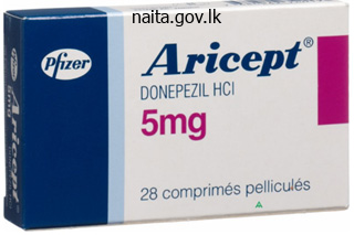
Order donepezil 10 mg free shipping
Interestingly, the cardiomyocytes have been of myocardial quite than of bone marrow origin, suggesting that resident cardiac stem cells exist and are stimulated by cytokines. These studies are promising; nevertheless, a mixture of cytokines had been used making it troublesome to elucidate the importance of stem cell issue alone. Ayach et al106 clarified the necessary contribution of stem cell think about cardiac repair in a study performed in c-kit poor mice. Moreover, Ayach et al showed that mobilization of natural killer cells was necessary in c-kit mediated cardiac repair. In summary, stem cell factor is a potent cytokine that induces mobilization of bone marrow�derived stem cells, angiogenesis, and recruitment of inflammatory cells. Animal studies have been promising, showing the helpful results of stem cell factor; however, most studies have looked at using stem cell factor together with different cytokines. Animal Studies Stem cell issue works in a wide range of methods to provide cardioprotection within the setting of ischemia, together with mobilization of bone marrow stem cells, stimulation of angiogenesis, proliferation of cardiomyocytes, reduction in apoptosis, and recruitment of mast cells. This could additionally be because of the inaccurate timing of cytokine administration, incorrect doses, or an insufficient homing effect of cytokines. In addition, a cocktail of cytokines may prove to be more beneficial than anybody cytokine alone. Intensive efforts over the past forty five years to develop essential pharmacologic therapies have decreased cardiovascular risk dramatically. A new calcium channel blocker was launched for the remedy of hypertensive emergencies (Chapter eight, Calcium Channel Blockers), as was a new antiarrhythmic for the remedy of arrhythmias (Chapter 17, Antiarrhythmic Drugs). An totally new class of drugs was launched for the treatment of angina pectoris (Chapter 15, Ranolazine: A Piperazine Derivative), and drug-eluting coronary stents may substitute naked metallic stents for patients with coronary artery illness (Chapter 29, Drug-Eluting Stents). There are currently over 100 pharmacologic treatments under investigation for the management of cardiovascular disease, lots of them encompassing new technologies and pharmacologic courses. Entire new lessons of agents are being studied in sufferers to stop atherosclerosis, which embrace phospholipase inhibitors;9,9a p38 kinase inhibitors; liver X agonists;10 niacin receptor agonists (see Table 37-8); inhibitors of toll-like receptors;11 and stimulators of heme oxygenase. Heart failure is a rising problem within the United States, and investigations utilizing new medication embody vasopressin antagonists,17 relaxin,18 direct renin inhibitors, adenosine agonists,19,20 cell-based therapy, and gene therapies. For the therapy of systemic hypertension, vaccines are in improvement,21 as well as new lessons of drugs20-24 and new combos of already approved agents (Table 37-7, Chapter 21, New Aspects of Combination Therapy: Focus on Hypertension). Innovative drug therapies that are being evaluated for peripheral vascular diseases (Table 37-10), pulmonary hypertension (Table 37-11), and cerebrovascular disease (Table 37-12) embrace stimulators of soluble guanylate cyclase25 and nitric oxide. In addition, numerous gene therapy therapy approaches and endothelial cell therapy26 are being studied (Tables 37-10, 37-11). These new therapies, lots of which had been developed out of recent scientific advances in molecular biology, molecular pharmacology, and translational analysis, show great promise in continuing the outstanding progress being made in opposition to cardiovascular disease and stroke. We predict that lots of the new treatments being discussed in this chapter could have become commonplace therapies by the point the fourth edition of this e-book is printed. Abciximab (c7E Fab): A review of its pharmacology and therapeutic potential in ischemic coronary heart disease. The pharmacokinetic profile of seventy four � 17 93 � 1 16 � 4 39 � eight 10 Cl is in eld and amlodipine. Atorvastatin: A evaluate of its pharmacology and therapeutic potential in the management of hyperlipidaemias. Intramuscular atropine in wholesome volunteers: A pharmacokinetic and pharmacodynamic research. Anticoagulant Bivalirudin 401 min exercise of Hirulog, a direct thrombin inhibitor, in humans. GenericName GenericName Clearance Therapeutic References (mL min-1 kg-1) Range 12. Captopril Carteolol - - Bioavailability Protein Volumeof Half-Life Urinary (%) Binding(%) Distribution (hours) Excretion (liters/kg) (%unchanged) 40�50 65�75 30 � 6 zero. Ocular carteolol: A review of its pharmacological properties, and therapeutic use in glaucoma and ocular hypertension.
Donepezil 10 mg with mastercard
The examination Once you decide to take the exam, set out about discovering the proper method to put together for it. Passing an examination requires the following: Preparation (acquiring knowledge) Consolidation (of the knowledge) Delivery (being in a place to express efficiently what you know). I would like to recommend that, in addition to your individual individual preparation, you determine a research group and attend courses. These difficulties are brought in to sharp focus when it comes to taking an examination, which entails gathering data and practising it Most of the work needed for the examination should be done alone Acquiring information 1. This consists of: a) Improving your examination techniques: for example, introducing yourself, taking a historical past about comorbidities and allergic reactions, correct exposure, asking the affected person to get up and walk and never forgetting to examine the patellofemoral joint in knee Postgraduate Orthopaedics, 2nd Edition, ed. Why am I prescribing medicine and how do antiinflammatories, antibiotics, native anaesthetics and steroids work If I am itemizing a affected person for surgical procedure, then I must be clear as to the indication, preoperative planning, exposure, postoperative administration, etc. Divide this in to two: Theoretical knowledge Work-based data Theoretical knowledge You need to learn a lot Read books of your alternative Visit various websites; significantly Orthoteers and Orthopaedic Hyperguide. Work-based information I knew my limitations regarding expertise in anything apart from general orthopaedics. I had worked only in a single hospital for 10 years and seriously lacked sensible experience in paediatric orthopaedics, hand and backbone. I arranged to meet with advisor paediatric orthopaedic, hand and spinal surgeons, and asked if I could have a clinical attachment with them in order that I could achieve some useful experience in these specialties pertaining to my exam. Having received their permission, I attended their clinics and theatres within the above specialties. In hand surgical procedure I discovered: Wrist examination Classification and management of swan neck and boutonni�re deformities Different hand splints Management of the rheumatoid hand Management of wrist instability. In spine I discovered: Examination of the backbone Principles of administration of spinal fractures Different approaches to the spine and spinal decompression. The issues you must ask the trainee are: What the trainee learned on their teaching sessions. Courses I would strongly advocate the course organized by the Royal College of Surgeons of England. Consolidation Facts may be acquired rapidly but they tend to fade away even quicker. This is a continuum of studying, remembering and practising the facts and is especially helpful for the clinical section of the examination. Therefore, I will inform my advisor as a result of: He/she can take me by way of lengthy and brief instances Give some time for oral practice Tell you his/her experiences, good and unhealthy May ask different advisor to assist you to May make certain your research leaves are permitted with out drawback. If you then method your marketing consultant with your intention this may be very unlikely that he/she might be discouraging. On the contrary, it is extremely possible that the marketing consultant will make an effort to polish off your knowledge earlier than the examination. What to do in case your consultants are unhelpful with either the form or teaching/practice, and so on. In the case that the advisor refuses to signal then I would do the next: Ask for his/her recommendation as to what I would have to do to change his/her mind Follow the recommendation and method again. If the response is unfavorable then I would: Ask one other advisor Speak to the medical lead, medical director, postgraduate tutor or intercollegiate board You will get there. These sessions are more likely to be brief however very useful and relevant to affected person administration. I have discovered that the easiest way to practise answering questions is to rehearse with colleagues in your research group. There follow the processes/practicalities of when you get your consultants to signal the form allowing you to sit the exam; tips on how to go about issues, etc. Before you ask a advisor to signal your advice form, mirror on these factors: Have you shown an interest in taking the exam these days The reception from the examiners within the oral and clinical was excellent and faultless. However, these days nearly all staff grades are nicely motivated and properly prepared, hence the cross rates are bettering. Consult the breakdown of the outcome sheet Identify the place and how the mistake was made Was it data that you lacked � then learn once more If it was a failure to convey the message, then practise it Listen to the question and answer that exact bit Relay the state of affairs to your consultant and ask his opinion Fill within the gaps in your information. It is extremely tough otherwise to show this degree of information and abilities. The analysis considers the coaching and/or skills, taking in to account knowledge and experience. The evidence to be collected shall be within the following domains: data, expertise and efficiency (75%), safety and quality (20%), communication, partnership and teamwork (5%) and sustaining trust (5%).
Buy cheap donepezil 10mg on line
Blockage of ventricular shunt catheters can end result in progressive ventricular dilation. Axial picture reveals high attenuation from acute hemorrhage inside dilated lateral ventricles. Axial picture shows dilated lateral ventricles and a porencephalic cyst on the right. Dilation of the left lateral ventricle secondary to encephalomalacia from old infarction within the vascular distribution of the left center cerebral artery. Dilation of the lateral ventricles in a neonate secondary to encephalomalacia from harmful changes of cerebritis. Axial picture shows uneven cerebral atrophy involving the frontal and temporal lobes with compensatory dilation of the ventricles. Axial image reveals uneven cerebral atrophy involving the frontal lobes with compensatory dilation of the ventricles. Occipital location commonest in Western hemisphere, frontoethmoidal location most typical website in Southeast Asians. Holoprosencephaly: Disorders of diverticulation (weeks 4�6 of gestation) characterised by absent or partial cleavage and differentiation of the embryonic cerebrum (prosencephalon) in to hemispheres and lobes. Semilobar: Monoventricle with partial formation of interhemispheric fissure, occipital and temporal horns, partially fused thalami. Fused inferior portions of frontal lobes, dysgenesis of corpus callosum, absence of septum pellucidum, separate thalami, neuronal migration problems. Septo-optic dysplasia (de Morsier syndrome): Mild form of lobar holoprosencephaly. Dysgenesis or agenesis of septum pellucidum, optic nerve hypoplasia, squared frontal horns; affiliation with schizencephaly in 50%. Absent or incomplete formation of gyri and sulci with shallow sylvian fissures and "determine 8" appearance of brain on axial images, abnormally thick cortex, gray matter heterotopia with easy gray-white matter interface. Associated with severe mental retardation, developmental delay, seizures, and early demise. Can have a bandlike (laminar) or nodular look isointense to grey matter; may be unilateral or bilateral. Neuronal migration dysfunction associated with hamartomatous overgrowth of the concerned hemisphere. I Intracranial Lesions Abnormal or Altered Configurations of the Ventricles 149 a. Axial photographs show fusion of the anteroinferior parts of the frontal lobes (a) with separation of the upper portions of the frontal lobes (b) with an interhemispheric fissure. Axial picture reveals nodular zones with intermediate attenuation along the margins of the lateral ventricles representing grey matter heterotopia. Axial picture reveals the absence of gyri and sulci and the dearth of regular gray-white matter demarcation. Axial image (a) exhibits open lip schizencephaly lined by gray matter alongside the margins. Axial picture (b) in a young baby with congenital toxoplasmosis with closed lip schizencephaly on the left, dystrophic calcifications at sites of prior an infection, and encephaloclastic modifications (arrow). Axial picture shows enlargement of the left cerebral hemisphere with irregular gyral configuration and zones of decreased attenuation in the left frontal lobe. Vermian aplasia or extreme hypoplasia; communication of fourth ventricle with retrocerebellar cyst; enlarged posterior fossa, high place of tentorium and transverse venous sinuses. Associated with other anomalies similar to dysgenesis of the corpus callosum, gray matter heterotopia, schizencephaly, holoprosencephaly, and cephaloceles. Spectrum of abnormalities starting from full to partial absence of the corpus callosum. Widely separated and parallel orientations of frontal horns and our bodies of lateral ventricles; excessive position of third ventricle in relation to interhemispheric fissure, colpocephaly. Comments Complex anomaly involving the cerebrum, cerebellum, brainstem, spinal twine, ventricles, cranium, and dura.
Donepezil 10 mg overnight delivery
The suprascapular nerve additionally sends some filaments to provide the shoulder joint and capsule. Descends posterior to clavicle and anterior to the brachial plexus and subclavian artery. May give a contribution to the phrenic nerve (C5), named the accent phrenic nerve if present. It arises obliquely behind the decrease fibres of pectoralis minor, lying lateral to the axillary artery and passes between the 2 conjoined heads of coracobrachialis. It runs laterally between biceps and brachialis adherent to the deep surface of biceps. It pierces the deep fascia, continuing on as the lateral cutaneous nerve of the forearm (lateral to the cephalic vein) supplying skin on the lateral facet of the forearm. The nerve is susceptible to damage during anterior dislocation of the shoulder owing to its close relationship with the inferior capsule. At the lower border of subscapularis it turns backwards and passes through the quadrangular area and then winds across the surgical neck of humerus with the posterior circumflex humeral vessels. After giving off a branch to the shoulder joint it divides in to anterior and posterior branches. The anterior department runs ahead around the humerus involved with the periosteum to enter the deep floor of deltoid. The posterior branch provides teres minor and winds across the posterior border of deltoid. It ends because the higher lateral cutaneous nerve of the arm supplying the skin over the inferior half of the deltoid. Radial nerve (C5, C6, C7, C8, T1): bigger terminal department of the posterior wire (largest department of the brachial plexus). It crosses the lower border of the posterior axillary wall, mendacity on latissimus dorsi and teres main. It passes by way of a triangular area below the decrease border of teres main, between the long head of triceps and the humerus. It spirals around the humerus (medial to lateral) within the spiral groove between medial and lateral heads of triceps along with the profunda brachii artery. It pierces the lateral intermuscular septum at the midpoint of the humerus to attain the anterior compartment between the brachialis and brachioradialis. It crosses the anterior aspect of the lateral epicondyle (where it provides anconeus) and enters the forearm, dividing in to deep and superficial branches. At the elbow the radial nerve lies on the elbow capsule on the midportion of the capitellum, making it vulnerable to injury during arthroscopic capsular release. Axilla Branches to long, medial and lateral heads of triceps Posterior cutaneous nerve of arm (posterior upper arm skin) Lower lateral cutaneous nerve of arm (lower lateral arm skin). Posterior compartment of the arm Motor to brachioradialis, brachialis (small provide, musculocutaneous main nerve), extensor carpi radialis longus Posterior cutaneous nerve of forearm (posterior forearm skin). Lower subscapular nerve (C6): provides the decrease part of subscapularis and teres major. It passes deep to brachialis proximal to the radial styloid and over the tendons of the snuffbox to attain the dorsal radial facet of the hand. Compression of the radial nerve at the elbow produces radial tunnel compression syndrome. The syndrome presents with forearm ache with out muscular weak spot and is commonly misdiagnosed as tennis elbow. Anterior interosseous nerve this arises slightly below the 2 heads of pronator teres to run on the interosseous membrane between flexor digitorum profundus and flexor pollicis longus to reach pronator quadratus. It supplies these muscles, except for the medial half of flexor digitorum profundus. Anterior interosseus nerve palsy presents principally as weak spot of the thumb and index finger. Palmar cutaneous branch of the median nerve this arises simply proximal to flexor retinaculum and turns into cutaneous between palmaris longus and flexor carpi radialis. It passes superficial to the flexor retinaculum to supply the lateral facet of the palm (skin). It helps to decide the level of a median nerve injury; numbness over the thenar eminence might point out a excessive lesion, while intact sensation with loss of operate in the recurrent and palmar digital branches could point out a more distal lesion.
Donepezil 10 mg cheap
Colonic interposition Various segments of the colon may be used to bypass lengthy segments of esophageal involvement by caustic strictures, superior achalasia, or inoperable esophageal cancers. This affected person has marked narrowing (arrow) at the pyloromyotomy web site because of postsurgical scarring in this region. Also observe dilatation of the stomach and retained particles despite the absence of any narrowing the place it traverses the diaphragm. These anastomotic strictures are often thought to develop as the sequelae of earlier leaks. In contrast, other patients could develop long, clean, relatively tapered non-anastomotic strictures of the interposed colon secondary to continual sixty eight Chapter three: Esophagus. Note how the intrathoracic abdomen flops inferiorly and to the best, delaying gastric emptying because of the effect of gravity. A research with water-soluble contrast materials exhibits small, sealed-off leaks (arrows) from each side of the proximal esophagocolic anastomosis. A quick section of clean, symmetric narrowing (arrow) is seen on the proximal esophagocolic anastomosis because of postsurgical scarring on this area. This affected person has an extended section of smooth narrowing (white arrows) with tapered margins (large black arrows) in the interposed colon distal to the esophagocolic anastomosis (small black arrow). These non-anastomotic strictures are thought to develop on account of chronic ischemia. Pneumatic dilatation and Heller myotomy Achalasia may be handled by pneumatic dilatation or by injection of the C. This focal, often eccentric ballooning (arrows) is assumed to end result from weakening of the wall of the distal esophagus on the site of the myotomy. The narrowed phase has abrupt, shelf-like distal margins (white arrows) due to tumor ingrowth through the uncovered distal end of the stent. Endoscopic biopsy specimens from this region revealed epithelial hyperplasia without evidence of tumor. The stent should be positioned with its proximal tip above the tumor and its distal tip below the tumor, and distinction materials should circulate freely via a broadly patent stent lumen. Even when a lined stent has been satisfactorily positioned for palliation of an esophageal-airway fistula, barium (and ingested liquid) may still enter the airway if it passes round quite than via the stent in to the fistula. Luminal narrowing and obstruction of the stent may be caused by tumor ingrowth through the stent wall. Stenting of tumors on the gastroesophageal junction is especially problematic due to massive reflux that usually occurs through the stent, causing nocturnal aspiration. In contrast, persistent dysphagia during the late postoperative interval could indicate an incomplete myotomy or a tight fundoplication wrap. Detection of gastroesophageal reflux: value of barium studies compared with 24-hr pH monitoring. Detection of reflux esophagitis on double-contrast esophagrams and endoscopy utilizing the histologic findings as the gold normal. Usefulness of barium research for differentiating benign and malignant strictures of the esophagus. Giant ulcers of the esophagus in sufferers with human immunodeficiency virus: clinical, radiographic, and pathologic findings. The small-caliber esophagus: radiographic signal of idiopathic eosinophilic esophagitis. Small benign tumors of the esophagus: radiological prognosis with double-contrast examination. Spindle-cell squamous carcinoma of the esophagus: a tumor with biphasic morphology. Primary malignant melanoma of the esophagus: radiographic findings in seven sufferers. Radiographic and endoscopic sensitivity in detecting decrease esophageal mucosal ring. Overlap phenomenon: a possible pitfall within the radiographic detection of decrease esophageal rings. Secondary achalasia and different esophageal motility disorders after Nissen fundoplication for gastroesophageal reflux disease.
Purchase donepezil cheap online
Axial picture exhibits bilateral zones of decreased attenuation involving the basal ganglia. Axial picture reveals a lesion with slightly increased attenuation in the left basal ganglia region containing several small calcifications (arrow). Axial images from three totally different sufferers show acute hematomas with high attenuation in the proper basal ganglia (a) and left basal ganglia (b,c) with various degrees of related mass effect. Atrophic modifications involving the mind can be seen later that could be related to cognitive impairment. Methanol intoxication Toxic results lead to selective necrosis of basal ganglia/putamina bilaterally and subcortical white matter 12 to 24 hours after ingestion secondary to metabolic conversion by hepatic alcohol dehydrogenase to the toxin formic acid. Mitochondrial disorder associated with external ophthalmoplegia, retinitis pigmentosa, and onset of medical muscular and neurologic signs in sufferers younger than 20 y. Wilson disease Autosomal recessive illness manifest by decreased useful serum ceruloplasmin ranges and altered copper metabolism with increased urinary excretion of copper. Usually presents in childhood with irregular poisonous copper deposition in tissues, resulting in cirrhosis and degenerative changes within the basal ganglia (lentiform nuclei) and brainstem. Axial image exhibits abnormal decreased attenuation within the globus pallidus regions bilaterally from necrosis. Axial picture shows calcifications within the basal ganglia areas and throughout the cerebral white matter bilaterally. Comments Autonomic dysfunction in adults with orthostatic hypotension; cerebellar and extrapyramidal clinical signs. Increased iron deposition and destruction of globus pallidus and substantia nigra bilaterally. X-linked (type 1) or autosomal recessive (type 2) leukodystrophy; 5 subtypes; deficiency of proteolipid element of myelin; presentation during neonatal period (type 2)/infancy (type 1) with irregular eye actions, nystagmus, delayed psychomotor development; death in first decade; males females. Slightly reduced diffusion in ventral thalamic nucleus, progressive cerebral and cerebellar atrophy. Tay-Sachs disease: functional hexosaminidase deficiency; neuronal ceroid-lipofuscinosis: lipofuscin deposits in cytosomes; mucopolysaccharidoses: autosomal recessive or X-linked issues associated to irregular metabolism of mucopolysaccharides (Hurler, Hunter, Sanfilippo, and Morquio syndromes). Result in axonal loss and demyelination related to the buildup of abnormal metabolites inside cells. Disorders of amino acid metabolism Autosomal recessive problems involving faulty enzymes regulating amino acid metabolism and mitochondrial operate. These enzymatic defects can cause vital alteration of regular formation and upkeep of myelin. Calcifications involving the basal ganglia in patients younger than or older than 30 y may be seen with hypoparathyroidism, pseudohypoparathyroidism, pseudopseudo-hypoparathyroidism, hyperparathyroidism, hypothyroidism, lead toxicity, Fahr disease, and neurodegeneration with mind iron accumulation. Comments Signal of calcium deposition can vary depending on the dimensions and configuration of deposits. Calcifications can also happen in basal ganglia secondary to endocrine abnormalities involving calcium (hypoparathyroidism), hypothyroidism, iron metabolism (neurodegeneration with brain iron accumulation), prior inflammatory disease (toxoplasmosis, tuberculosis, cysticercosis, and so on. Signal abnormality related to hepatic dysfunction (alcoholic cirrhosis, hepatitis, portal-systemic shunts) probably related to increased serum ammonia and manganese levels. Usually presents after age forty y with progressive movement issues (choreoathetosis, rigidity, hypokinesia); behavioral and progressive mental dysfunction/dementia. Juvenile Huntington disease also occurs in a small number of sufferers within the second decade. Patients current with rigidity, hypokinesia, seizures, and/or progressive mental dysfunction. Axial picture reveals zones of decreased attenuation in the thalami and to a lesser extent within the basal ganglia. Axial picture reveals bilateral calcifications in the basal ganglia, thalami, and cerebral white matter. Severe dysfunction of neuronal migration (weeks 7�16 of gestation) with absent or incomplete formation of gyri, sulci, and sylvian fissures. Pachygyria (nonlissencephalic cortical dysplasia) Gray matter heterotopia Thick gyri with shallow sulci involving all or parts of the brain. Thickened cortex with comparatively smooth graywhite interface may have areas of decreased attenuation within the white matter (gliosis). Laminar heterotopia appears as a band or bands of gray matter within the cerebral white matter.
Diseases
- Goniodysgenesis mental retardation short stature
- Spinocerebellar ataxia amyotrophy deafness
- Marshall Smith syndrome
- Uniparental disomy of 6
- Congenital giant megaureter
- Pulmonary surfactant protein B, deficiency of
Order 10 mg donepezil visa
Complications Re-fracture or non-union Stiffness of ankle and subtalar joints Limb shortening Progressive anterior angulation of tibia Infection Repeated operations Soft-tissue scarring. Congenital pseudoarthrosis of the tibia this can be a uncommon condition, with an incidence of 1:250 000. It presents with a spectrum of issues, starting from anterolateral bowing to frank pseudoarthrosis or pathological fracture with an apex deformity. Fibular hemimelia Definition this situation consists of a spectrum of anomalies from mild fibular shortening to whole absence of the fibula. Achterman and Kalamchi24 Type I: hypoplastic fibula Type Ia: proximal fibular epiphysis is more distal than normal, and distal fibular epiphysis is more proximal than normal. Angular deformities of the tibia are common and are related to severe foot and ankle problems (tarsal coalition, lateral ray deficiencies). Clinical features the concerned leg is short with a varus or calcaneovarus foot There is commonly a pores and skin dimple over the front of the leg Quadriceps muscle is commonly underdeveloped or absent; there are numerous levels of fixed flexion at the knee. Management Reconstruction options these embody: Distal fibulotalar arthrodesis or calcaneal�fibula fusion to stabilize the hindfoot Tibiofibular synostosis (fusion) Tibial lengthening with epiphysiodesis of the ipsilateral distal fibula and contralateral limb. Generally, the next ideas apply in deciding on reconstruction versus amputation: Mild deformity: reconstruct Severe: amputate Intermediate: get hold of a second opinion. Reconstruction choices include: Posterolateral launch to appropriate equinovalgus deformity of the foot Limb lengthening is indicated if the foot and ankle are comparatively regular. Avoid above- or below-diaphyseal amputations due to related problems with overgrowth of the residual diaphysis. Popliteal cyst the common site is medial, originating in the gastrocnemius� semimembranosus bursa just under the popliteal crease. The cyst arises from the synovial sheaths of the encircling tendons and accommodates clear viscous fluid. Tibial hemimelia Definition this condition represents a spectrum of deformities ranging from whole absence of the tibia to gentle hypoplasia. There are only a few indications for surgery: When the diagnosis is unsure Severe pain (check for different, extra apparent, causes) Sinister trigger. Examination nook Paeds oral: Clinical photograph of a kid with obvious swelling at the again of the knee Spot analysis with discussion afterwards of management, significantly the way you deal with awkward parents demanding surgery for his or her baby (second opinion! The swelling appears to be about three cm by 2 cm; the skin overlying the swelling seems normal. Estimate the angle made by an imaginary straight line along the axis of the thigh and an imaginary line along the axis of the foot. Transmalleolar�thigh angle the transmalleolar axis is marked by palpating the medial and lateral malleoli joining the two factors on the heel. A line perpendicular to this axis and the longitudinal axis of the thigh is assessed. Range of hip rotation (Staheli) Place the child in the inclined place with their knee flexed at 90� and their ankle held in neutral position. Clinical approach In-toeing accounts for the best variety of paediatric orthopaedic referrals in the developed world. The natural history of most instances of in-toeing (owing to extensive normal variability in femoral anteversion) is of gradual spontaneous resolution over the first 7 or 8 years of life. Flex the knees to 90� and internally rotate the leg while palpating the higher trochanter. Obstetric brachial plexus palsy that is stated to happen in a single in 2000 stay births, with 75% of cases totally recovering, although these figures are extremely topic to case ascertainment bias. The assessment of an older child with late presenting obstetric brachial plexus palsy has been seen in the examination. It is a difficult and extremely specialized space of apply, but you should nonetheless have an appreciation of the situation and its consequences. Differential diagnosis Pseudoparalysis owing to isolated clavicular fracture (which improves within days) Arthrogryposis Cerebral palsy. Natural history Aetiology Function spontaneously recovers generally (neurapraxia), normally distally first Residual issues typically contain the shoulder Biceps restoration before 2 months of age is an efficient prognostic sign; conversely, failure of biceps to recuperate by 4 months is a poor prognosticator. The trigger is shoulder dystocia � in a cephalic delivery the shoulders are too wide for the pelvis and the upper roots are stretched to ship the toddler, whereas in breech supply the decrease roots are extra weak to being stretched. Risk factors embrace giant start weight, instrumented supply, C-section, breech. The concern of the method to K-wire a supracondylar fracture has generated disproportionate consideration.
Purchase donepezil 5 mg amex
Open fractures and open joints are limbthreatening and wish irrigation, tetanus immunization, and parenteral antibiotics. Traumatic amputations will want to be thought-about for emergency reimplantation, and the amputated half appropriately cooled and carefully cared for. Compartment syndrome might develop over hours, and thus high-risk accidents want serial bodily examinations and compartment pressures evaluated. Dislocations need immediate reduction with deep sedation and neurovascular status checked earlier than and after the discount. Closed fractures want at least gross alignment with splinting within the emergency division to decrease bleeding and pain and prevent further displacement and injury of the adjacent neurovascular constructions. Definitive closed or open reduction may be completed once the affected person has been stabilized from different accidents. Significant occult blood loss can happen with fractures of enormous bones such as the femur and pelvis and may typically account for acute hemorrhagic shock. Anticipate massive blood loss with these injuries while excluding different causes of hemorrhage, 2. Compartment syndrome might insidiously develop within the multitrauma patient, and thus repeat medical examination is paramount for early prognosis of this situation. Pain out of proportion to the obvious damage is an early symptom and will need to be evaluated by assessing compartment pressures. Segmental fractures in long bones such because the tibia are particularly prone to compartment syndrome. Occult fractures must be suspected in sufferers with important pain but with regular radiographs. Pediatric radiographs are inherently tougher to interpret because of osseous growth plates and incomplete ossification. At instances, comparability views of the other extremity shall be needed, and occult fractures ought to be suspected in kids with tenderness over the physis. Long bone fractures should be splinted before transferring the affected person to the radiology suite. Immobilization of the fracture reduces ache and bleeding and prevents additional harm to the neurovascular structures. Fractures are first described by anatomic location, and lengthy bones are often divided in thirds describing the location of the damage. Open or compound fractures check with fractures with a break within the overlying pores and skin, in contrast to closed fractures which have regular skin integrity. The fracture line or pattern is then described and follows the following widespread conference: 1. Displacement refers to the diploma of offset of the bone ends relative to each other, and completely displaced fractures are inclined to be extra unstable accidents. Displacement is described by outlining the position of the distal bone relative to the proximal end. A bayonet deformity refers to injuries with 100% displacement and overriding of the bone ends with shortening of the affected extremity. Angulation refers to the angle between the longitudinal axes of the principle fracture segments. Fractures with significant angulation typically require reduction to maintain good function. They are outlined as fractures in touch with the skin environment and thus require a break in the pores and skin covering the fracture website. The skin break may be large and obvious or a small puncture wound damage, and thus dedication if a fracture is open can generally be tough. The Gustilo classification is often used when describing open fractures: Type I: Puncture wound <1 cm and comparatively clear. Emergency remedy consists of overlaying the wound with sterile saline dressings, along with the suitable tetanus immunization and ache management. Patients needing emergent operation for extreme associated accidents can have external fixation performed whereas stable patients are eligible for inner fixation. There is a mix of orthopedic trauma, in depth delicate tissue harm, and injury to the neurovascular constructions.
Donepezil 5mg without prescription
Because of the stereotopic organization of the lateral corticospinal tract, the palms (lying closest to the center of the cord) are extra affected than the arms, which in turn are extra affected than the lower extremities. Plain radiographs of the cervical spine are often normal because the condition could happen with no fracture or dislocation. Misdiagnosis of the condition as "malingering" or "conversion dysfunction" is widespread. Treatment is immobilization of the neck and administration of high-dose corticosteroids. Recovery of bowel and bladder perform and ambulation is the rule, though restoration of full guide dexterity is uncommon. It consists of a attribute constellation of findings with loss of pain and temperature contralateral to the side of the damage and lack of motor function (corticospinal tract), mild touch (spinothalamic tract), position, and vibration sensation (posterior columns) ipsilateral to the harm. Broad-spectrum antibiotic coverage is indicated, and surgical debridement may be required. Functional recovery is usually surprisingly good despite the everlasting twine lesions. Anterior cord syndrome is often seen in aged patients along side medical situations, notably arterial embolization from the center. However, it may additionally result from prolonged cross clamping of the aorta, prolonged hypotension, or direct harm from bone fragments or international our bodies. Upper Cervical Spine Dislocations There are multiple sturdy ligamentous attachments from the cervical backbone to the base of the skull. These include the anterior and posterior longitudinal ligaments, the anterior and posterior atlanto-occipital membranes, the apical ligament from the odontoid, and the capsular ligaments of the atlanto-occipital joint. Separation of the cervical backbone from the base of the skull is type of invariably fatal on the scene of the trauma. Fatalities happen from disruption of the spinal wire above 216 Spinal Injuries the level of C-4 which leads to complete paralysis of respiratory effort and asphyxial death or from injury to the brainstem or vertebral arteries. In sufferers who survive to attain a hospital, mortality is excessive because of brainstem damage, associated head and systemic trauma, and prolonged coma with its attendant complications. Nevertheless, survivors have been reported, and a full resuscitative effort ought to be made. The harm is primarily ligamentous, although associated fractures of the face and skull are widespread. Neurologic deficits are unusual with superior dislocation (as shown) or when associated with fracture of the odontoid, although the situation is unstable because of disruption of the ligaments. Anterior dislocation is normally fatal because the spinal cord is compressed in opposition to the intact odontoid. The ring of C-1 (A) is seen mendacity utterly above the tip of the odontoid course of (B). The plain radiographic look of this injury is refined and could additionally be confused with a burst fracture of C-1. In the case of rotatory subluxation, one lateral mass rotates anteriorly and the other posteriorly. Conversely, the right lateral mass will seem smaller and farther from the odontoid process. The proper lateral mass is seen clearly but the left lateral mass of C-1 has rotated posteriorly out of view. Rotary Subluxation C-1/C-2 H nearer to the odontoid process, whereas the opposite lateral mass appears smaller and farther from the odontoid. On the plain radiographic open-mouth odontoid view, the gap from the odontoid to the lateral mass is increased on one facet and decreased on the other, in contrast to a burst fracture of C-1, in which each lateral masses are displaced laterally. In burst fractures that occur on just one facet of the ring of C-1, that lateral mass will be displaced laterally, whereas the unaffected facet will present a standard relationship of the lateral mass of C-1 to the articular plate of C-2. They are contraindicated in sufferers with apparent neurologic harm or in these whose prognosis is obvious. The extension view seems regular, however the flexion view exhibits significant angulation and anterior subluxation of C-4 on C-5, indicating posterior ligamentous disruption.
Cheap 10 mg donepezil otc
Deep surgical dissection Split the longus colli muscle tissue, which lie on the anterior surface of the vertebral bodies. Incision Generous straight midline neck incision via pores and skin and subcutaneous fascia. Incision A longitudinal midline incision is made from just below the umbilicus to simply above the pubic symphysis. The incision is extended superiorly, curving it simply to the left of the umbilicus. Superficial surgical dissection the wound is deepened consistent with the skin incision right down to the spinous processes by incising via fascia and the nuchal ligament. Cobb elevators are used to clear the ideas of the spinous processes, and then the elevators are reversed and swept laterally to clear muscle attachments from the lamina (paraspinal muscles). Using deep self-retaining retractors, carry dissection as far laterally as essential to expose the lamina, facet joints and beginnings of the transverse processes. Internervous aircraft the midline aircraft lies between the abdominal muscular tissues on both sides, segmentally equipped by branches from the seventh to the twelfth intercostal nerves. Superficial surgical dissection the wound is deepened consistent with the pores and skin incision, chopping through fats to reach the fibrous rectus sheath. The two rectus abdominis muscle tissue are separated utilizing finger dissection to expose the peritoneum. The incision is extended proximally and distally, with one hand contained in the stomach cavity to shield the viscera. Deep surgical dissection Identify the ligamentum flavum that runs between the laminae. Using a pointy blade or Cobb elevator remove the ligamentum flavum from the vanguard of the lamina of the inferior vertebrae. Place a flat-shaped spatula within the midline between the ligamentum flavum and underlying dura. Perform a laminectomy (partial or complete), eradicating as much of the lamina as necessary to see the blue�white dura that lies instantly beneath it coated by epidural fats. Deep surgical dissection Insert a self-retaining retractor to retract the rectus abdominis muscular tissues away laterally. This is carried out by incising the peritoneum on the left side of the aorta, and mobilizing the colon and left ureter. Structures in danger Spinal cord and nerve roots Venous plexus within the cervical canal Vertebral artery. Anatomy of the paracervical muscular tissues the paracervical muscle tissue within the cervical spine run in three layers. The middle portion consists of semispinalis cervicis and the deep layer consists of the multifidus muscles and the brief and lengthy rotator muscle tissue. Structures at risk Middle sacral artery Aorta Inferior vena cava Presacral plexus of parasympathetic nerves (important for sexual function) Ureter Left lumbar vessels. Anterior approach to the lumbar backbone Position Supine on operating desk Urinary catheter Nasogastric tube. Position Kneeling place with flexion hips and knees to flex spine and open up interspinous areas (90/90 position) Or prone on cushions Lateral with affected side uppermost It is essential to reduce venous plexus filling around the spinal twine by allowing the venous plexus to drain immediately in to the inferior vena cava. The method could be extended proximally or distally, detaching the posterior spinal musculature from the posterior spinal elements as required. To gain higher exposure domestically to visualize the dura, nerve root or disc better, additional parts of the lamina could be eliminated. The venous plexus surrounding the nerves and the ground of the vertebra may bleed during the blunt dissection wanted to attain the disc. The iliac vessels mendacity on the anterior side of the vertebral our bodies could additionally be injured if instruments pass through the anterior portion of the annulus fibrosus. Landmarks Iliac crest (L4 spinous process) Radiographs with needle on the appropriate disc stage Spinous processes.
References
- Ramakrishna G, Sprung J, Ravi BS, et al: Impact of pulmonary hypertension on the outcomes of noncardiac surgery: Predictors of perioperative morbidity and mortality, J Am Coll Cardiol 45:1691- 1699, 2005.
- McMurray BR, Wrenn KD, Wright SW: Usefulness of blood cultures in pyelonephritis. Am J Emerg Med 15:137-140, 1997.
- Arbeit RD, Maki D, Tally FP, et al. The safety and effi cacy of daptomycin for the treatment of complicated skin and skin-structure infections. Clin Infect Dis. 2004;38:1673-1681.
- White, W.M., Haber, G.P., Goel, R.K., Crouzet, S., Stein, R.J., Kaouk, J.H. Single-port urological surgery: single-center experience with the first 100 cases. Urology 2009;74:801-804.
- Govindarajan A, Reidy D, Weiser MR, et al. Recurrence rates and prognostic factors in ypN0 rectal cancer after neoadjuvant chemoradiation and total mesorectal excision. Ann Surg Oncol 2011;18(13):3666-3672.
- Reichelt A, Hoeper MM, Laganski M, et al: Chronic thromboembolic pulmonary hypertension: Evaluation with 64-detector row CT versus digital subtraction angiography, Eur J Radiol 71:49, 2009.